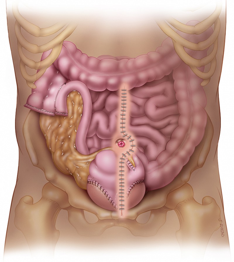
What is bladder augmentation? Bladder augmentation is increasing the size of the bladder, usually with a patch of bowel. This allows patients to store usually a lot of urine in their bladder. It is usually used for patients that have neurologic injury or disease that has caused the bladder to shrink and frequently spasm leading to leakage of urine.
How does increasing the size of the bladder with an augmentation surgery decrease leakage? Most neurologic disease causes the bladder to shrink and also to spasm. The low volume and high pressure of the spasms causes urine to leak through the sphincter muscles. Often the sphincter muscles are very strong, but the bladder pressure overpowers them and leakage results. When the bladder volume is increased the bladder can hold much more volume and this eliminates the spasms and the leakage.
What are the reasons that a bladder augmentation might be recommended? The typical reasons that your surgeon might recommend this surgery is when the bladder becomes very small from problems like spinal cord injury. Another very common scenario is when patients have physical limitations and cannot catheterize their urethra. A common situation like this is a woman with paraplegia that has to move out of her wheelchair in order to catheterize. This can be very difficult and limit a patient’s ability to travel and participate in life. In this situation the bladder is usually increased in size and at the same time a channel of small bowel is created that travels from the top of the bladder to the belly button (umbilicus). Then the patient can just lift up the lower portion of their shirt and pass the catheter from the belly button down to the bladder and drain out the bladder without changing positions. Another common scenario is in a patient with quadriplegia who has difficulty with passage of a catheter down the urethra. In order to catheterize, a patient must have good enough hand function to undue their zipper and pull their pants down to access the penis or the vagina. This combined with the difficulty of finding or passing the catheter down the urethra can be very challenging for someone with limited strength and hand function. When a catheterizable channel is brought up to the belly button, catheterization is much simpler and most quadriplegic patients that can write and pick up a pencil from a table can successfully catheterize these types of channels. A catheterizable channel can allow patients to be free of permanent catheters like a Foley or suprapubic catheter and successful do intermittent catheterization.
What are the different types of catheterizable channels and when does a patient need one of these channels? A catheterizable channel is a bowel tube made from appendix or small bowel that comes up to skin of the abdomen, often in the base of the belly button and connects to the bladder. If patients have trouble with catheterizing their urethra, these channels can help them successfully remain on self-catheterization, rather than going to a permanent catheter. There are a variety of ways of making these channels depending upon whether patients need to have the bladder expanded or just need to have the channel made for ease of catheterization.
What is a small bowel augmentation surgery? In this surgery the bladder volume is increased with a U of small bowel that is opened and made into a rectangle. The rectangle is sewn to the bladder and increases its volume dramatically. In this surgery a separate catheterization channel is not created and patients still catheterize their urethra.
What is the surgery cutaneous catheterizable ileocecocystoplasty? This is a surgery that we have worked hard to develop at University of Utah and we have found to be very successful. We encourage you to view the video on this topic. In this surgery the bladder is expanded with large bowel from the cecum and ascending colon. The connection to the small bowel serves as the catheterizable channel, which comes up to the belly button. This small bowel (terminal ileum) is narrowed to about the diameter of a pencil. There is a natural valve (ileocecal valve) between the small bowel and large bowel that prevents leakage out of the channel. The surgery both increases the size of the bladder and creates a catheterizable channel when patients cannot easily catheterize the urethra.
What is involved in surgery? The surgery takes about 5-6 hours to perform. Patients usually are in the hospital after surgery about 7-14 days. The main thing that keeps them in the hospital is the return of the bowels to proper function. It takes a while for patients to begin to eat and have normal bowel movements after a piece of the bowel is used to reconstruct the bladder. Patients then have a large suprapubic tube that drains the bladder while it heals. This stays in place for 1 month then patients begin to catheterize the bladder either via their urethra or from the catheterizable channel. The suprapubic tube is removed after patients are catheterizing successfully without problems, usually a couple of weeks later. At first patients catheterize frequently, but time is added in between catheterizations until they are catheterizing about 4-5 times in 24 hours.
What are some of the complications associated with the surgery? There are some immediate complications from surgery that we watch for very closely. These are problems like bowel obstruction, hernia, bowel or bladder fistula, and post-operative infections. The long-term complications can be problems like leakage from the catheterizable channel, or difficulty with catheterization. These problems occur in about 10% of patients and these patients may need revision of their catheterizable channel.
What is the post op recovery from the surgery? Bladder augmentation surgery is large abdominal surgery and it takes some time to recover. Patients need about 6 weeks until they begin to do their regular activities and may have some pain and healing that occurs up to 3 months after surgery. It is important to do no heavy lifting for about 6 weeks after surgery to prevent a hernia.
Figure 1 – The incision for augmentation of the bladder and creation of a catheterizable channel is from the pubic bone to above the belly button (umbilicus).
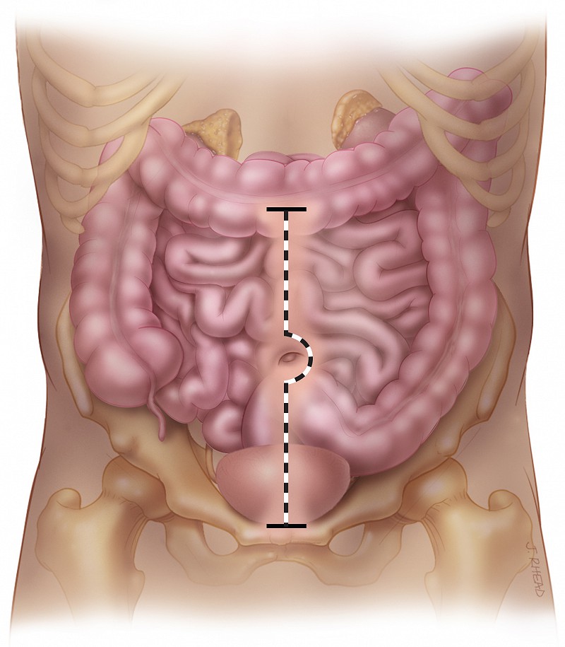
Figure 2 – The bladder is opened from front to back. This creates space for the bowel to be sewn in place and also acts to defunctionalize the bladder so it cannot spasm.
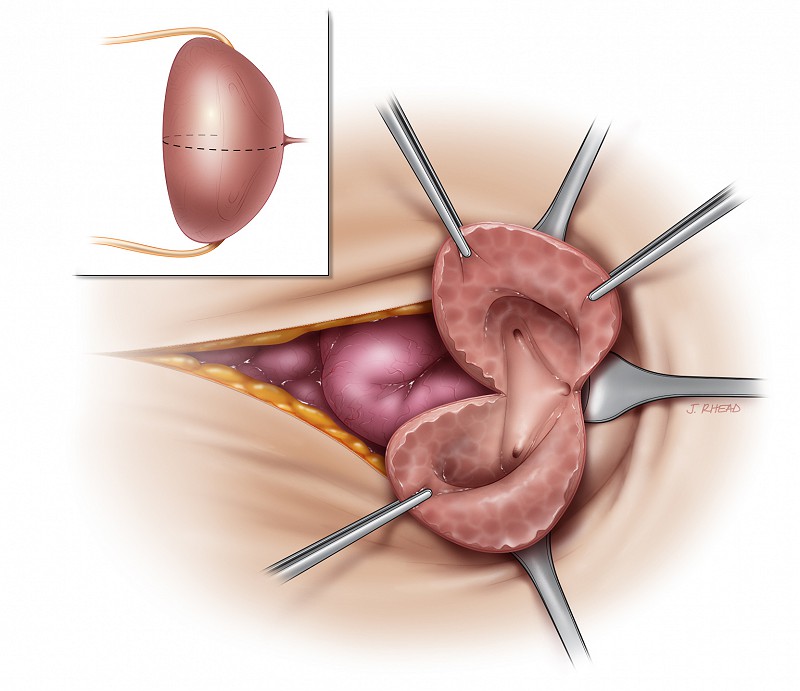
Figure 3 – The cecum, ascending colon, and terminal ileum is isolated to create the bowel segment for augmentation of the bladder and creation of the catheterizable channel.
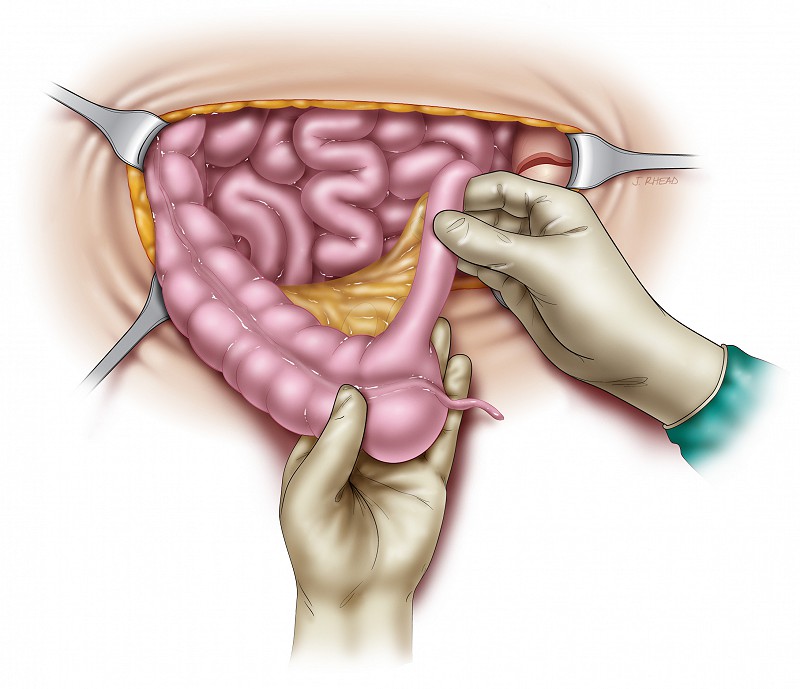
Figure 4 – The cecum, ascending colon and terminal ileum is detached from the bowel and the small bowel is reconnected to the large bowel.
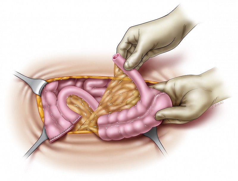
Figure 5 – The large bowel is opened to use for augmentation and the terminal ileum is reduced and tightened to serve as the catheterizable channel.
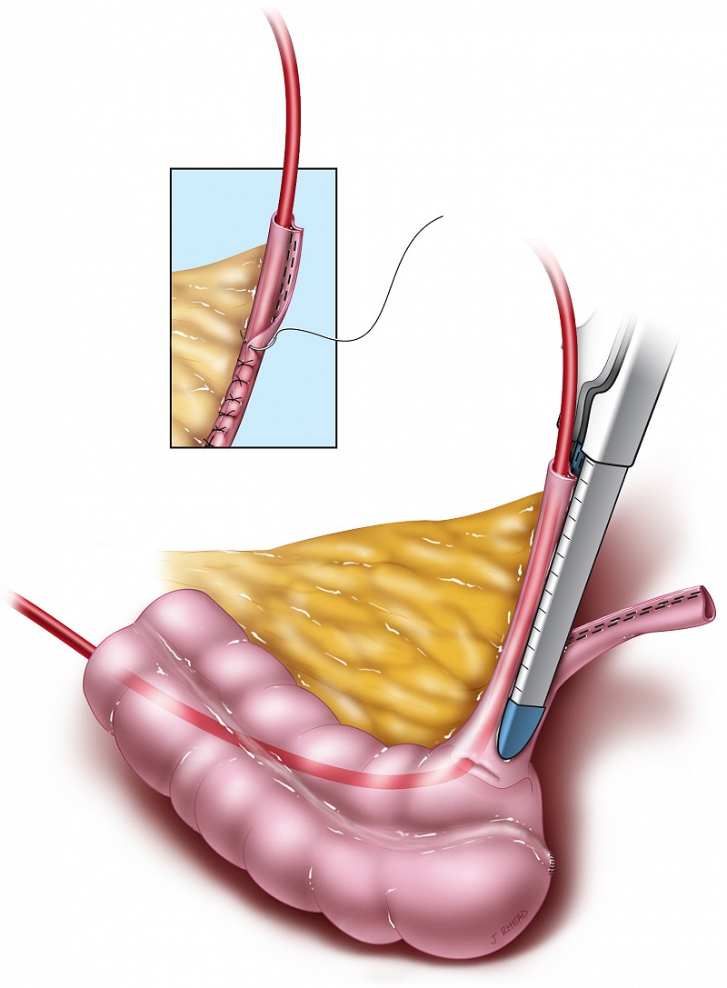
Figure 6 – Sewing the augmentation to the bladder to expand its volume.
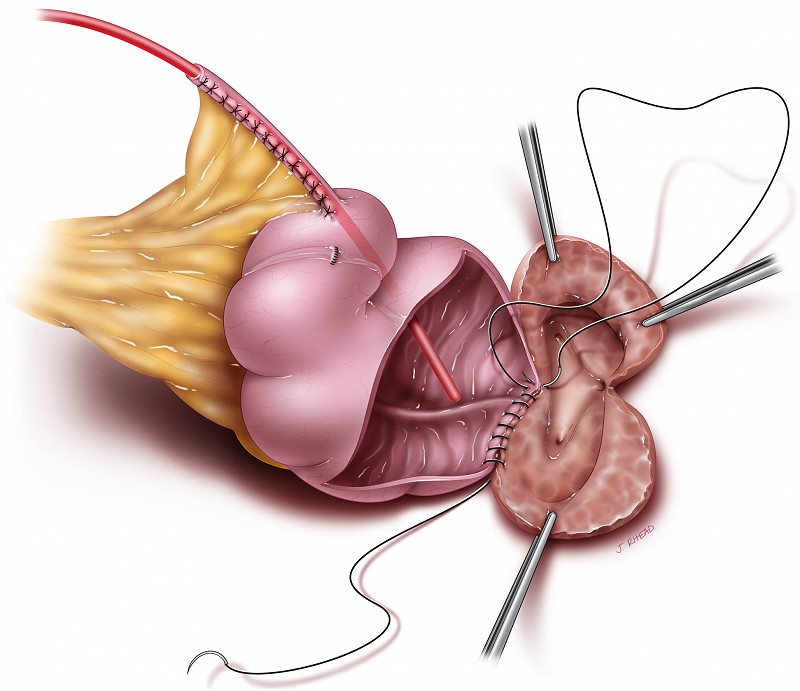
Figure 7 – After the augmentation of the bladder and creation of the catheterizable channel.
